Explore our reagents available with the fluorochrome BV421.
Multicolor Phenotyping of Antigen-Specific T Cells Using Immune Cell Profiling Panels Including MHC Dextramer® BV421
Deep Characterization of Circulating CMV-Specific T Cells
The design of many commercial panels for immunophenotyping accommodates the addition of BV421 labeled components. The aim of this study was to demonstrate that MHC Dextramer® BV421 reagents integrate seamlessly as components in commercial immune cell profiling panels to extend the analysis of surface markers to include information about antigen specificity. Multiparametric flow cytometry enables more comprehensive T cell monitoring and a deeper characterization of T cell populations.
Immune Cell Profiling Panel
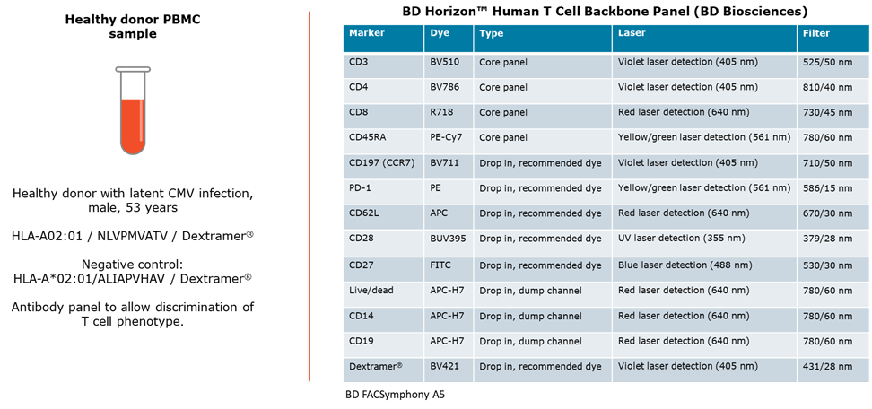
For this experiment we used the “BD Horizon™ Human T Cell Backbone Panel” from BD Biosciences. This panel includes 5 core T cell markers and 5 drop-ins on recommended fluorochromes. We used MHC Dextramer® BV421 as one of the added components along with four other relevant markers for phenotypic characterization of CMV-specific T cells in the blood of a donor with latent CMV infection.
Materials and Methods
The experiment was carried out following the MHC Dextramer® Staining Protocol (see immudex.com/resources/protocols).
After MHC Dextramer® and antibody staining, the samples were fixed using 2% BD Cytofix™ Fixation Buffer to preserve the cells until flow cytometric analysis was performed.
We utilized a BD FACSymphony™ A5 flow cytometer for sample analysis.
- Sample volume 130 µL
- Flow rate 1.5 µL/sec
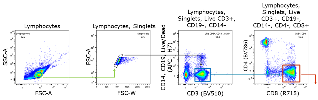
Gating Strategy
The PBMC sample was stained with the immune profiling panel and analyzed by flow cytometry. Cells were first gated as shown below. Successive subpopulations were then analyzed with the remaining markers as shown.
High T Cell Response in Latent CMV Infection

We observed a high T-cell response to CMV in the PBMC sample. CMV-specific T cells make up 11% of all CD8+ T cells, which is common in healthy individuals with latent infection.
It is a feature of CMV infections that T-cell responses do not contract after primary infection and that the number of CMV-specific cells remains high and even increases with age. This is a phenomenon known as “memory T cell inflation”.
T Cell Subsets Defined by Expression of Two Surface Molecules: CD45RA and CCR7
T cells can be divided into four functionally distinct subsets defined by the expression of CD45RA and CCR7 (Ref. 1).

CMV-specific T Cells are Predominantly of the TEMRA Phenotype with Downregulated Expression of Co-stimulatory Molecules CD27 & CD28
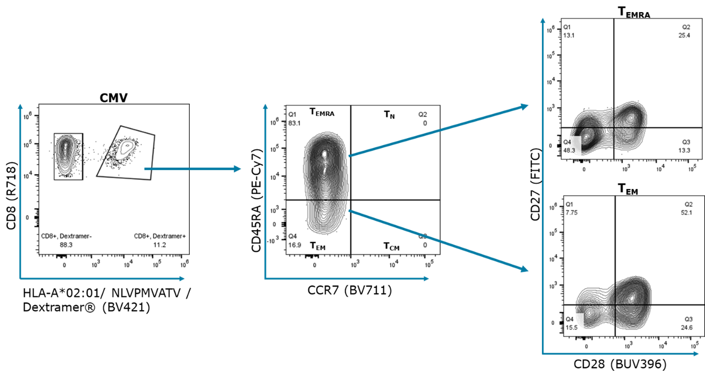
Circulating memory T cells are generally predominantly of the TEMRA phenotype. But this bias is known to be even stronger with CMV-specific T cells. In the tested individual CMV-specific TEMRA make up 83% of the memory T cells.
CMV-specific Memory Cells are Negative for Homing Receptors for Lymphoid Organs
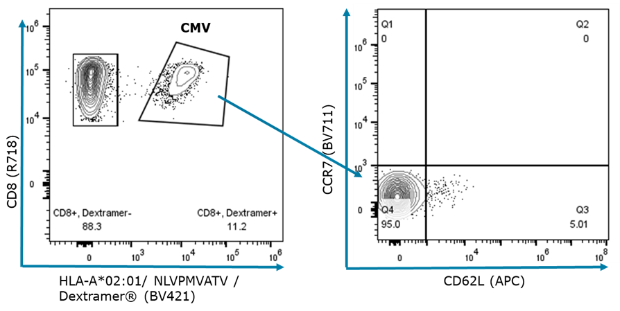
L-selectin (CD62L) and C-C chemokine receptor type 7 (CCR7) are essential for T cell migration to lymph nodes. As observed above CMV specific memory cells are largely negative for both proteins, which allows them to circulate throughout the body in peripheral tissue, spleen and blood.
Previously considered terminally differentiated TEMRA cells retain some level of plasticity. Evidence suggests that these memory cells upon antigen stimulation (by CMV reactivation) and multiple cell divisions later lose CD45RA and become CCR7+ allowing them to infiltrate lymph nodes again. Once the infection has come under control and after a long absence of antigen, the cells revert to the resting TEMRA phenotype (Ref. 2).
Most CMV-specific Memory Cells Express PD-1
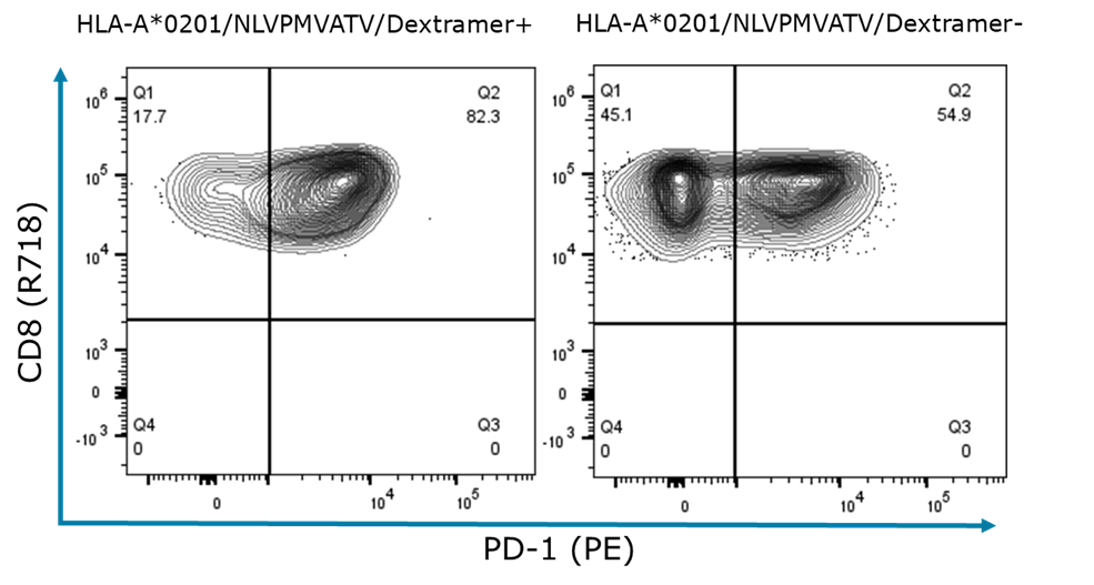
Most of the CMV-specific memory T cells express PD-1 and are in an activated state ready to pounce if the virus reactivates.
Conclusions
The study shows that MHC Dextramer® BV421 is easy to incorporate as an additional component into an existing T cell immune profiling panel.
Including the MHC Dextramer® reagents made it possible to obtain detailed information on CMV-specific T memory cells in blood from an individual with a dormant CMV infection.
References
¹ Sallusto, F. et al. Two subsets of memory T lymphocytes with distinct homing potentials and effector functions. Nature 401, 708–712 (1999). https://doi.org/10.1038/44385
² Carrasco J. et al. CD45RA on human CD8 T cells is sensitive to the time elapsed since the last antigenic stimulation. Blood, 108, 2897-2905 (2006). https://doi.org/10.1182/blood-2005-11-007237
Related Resources
Interested in Using MHC Dextramer® in Multicolor Flow Cytometry?
Fill in the form and one of our dedicated specialists will get in touch with you shortly.
BV421 is equivalent to Brilliant Violet™ 421, which is a trademark or registered trademark of Becton, Dickinson and Company or its affiliates, and is used under license. Powered by BD Innovation.